October 15th– World Anatomy day
In celebration of World Anatomy Day, we’re ‘dissecting’ an interesting book found in Raby’s collections.
Andreas Vesalius
World Anatomy Day honours the work of Andreas Vesalius on the anniversary of his death in 1564. Vesalius is considered the founder of human anatomy studies, through the creation of his seven-volume book De Humani Corporis Fabrica (on the fabric of the human body.) The work dissects the human body and considers each layer separately, from bones and cartilages, to ligaments and muscles, and the heart and other organs. The information is accompanied by classical illustrations and backgrounds of Italianate landscapes.
Published in 1543, the work was paired with a companion piece called the Epitome. This contained a brief summary of the anatomical structures in the Fabrica, but more interestingly a series of woodcuts of the dissected human body. This included a sheet which could be cut up and glued together by the reader to make a layered paper manikin (a medical model of the human body.) This helped readers- often medical students- to better understand how the human body was dissected and put together.
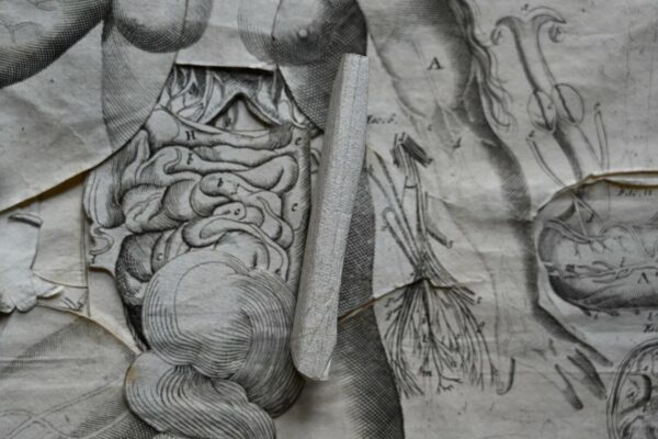
The book in Raby’s collection is similar in use and purpose to this Epitome by Vesalius.
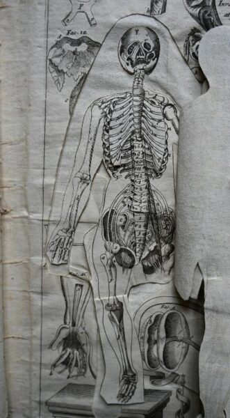
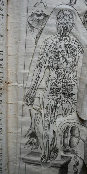
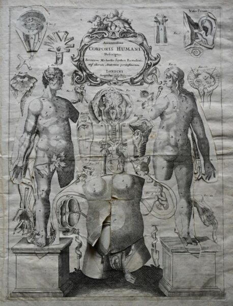
A section of the book showing the different layers of the body.
(Raby Collection)
Multi-layered anatomy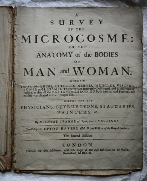
Illustrating anatomy using multi-layered flaps became popular during the sixteenth and seventeenth centuries. These were known as fugitive sheets, and were made up of engraved figures with additional sections as ‘flaps’ on different parts of the body. When lifted, these layers revealed illustrations of organs, blood vessels and bones.
This tradition began with Heinrich Vogtherr, who depicted a seated woman in 1538. A flap on the woman’s belly could be lifted to reveal her internal organs. He used the same woodblocks to create a male print a year later, changing the head and torso. Within the year, printers in other cities had created their own versions which were circulated throughout Europe.
They proved so popular that versions were created in several different languages, making specialist anatomic information more accessible to a wider audience.
The book in the Raby collection, ‘A Survey of the Microcosm, or the Anatomy of the Bodies of Man and Woman,’ is a good example of this, being an English translation of an earlier work. The sub heading explains that the book is ‘useful for all physicians, chyurgeons, statuaries, painters,’ showing its appeal to a wider audience than just those in the medical field.
Catoptrum Microcosmicum
The author of the original work from Raby’s collection was Johann Remmelin (1583-1632)- Remilinus in our English language version- a German medical doctor and anatomist. His Catoptrum Microcosmicum was first published in 1619 in Latin. It went through several editions in Latin, German, French, Dutch and English.
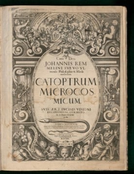
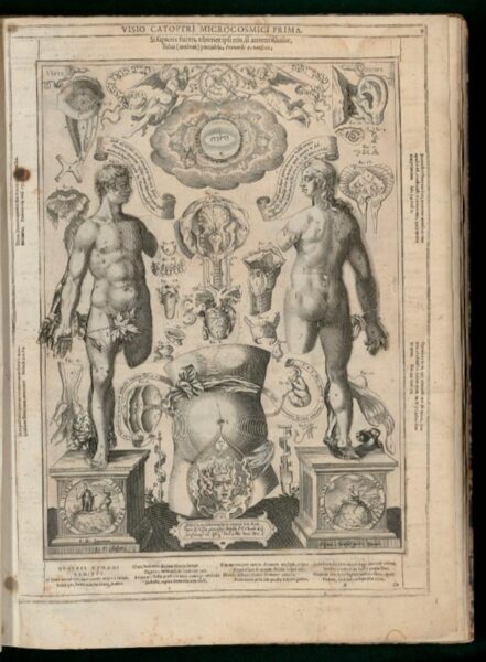
An original version of Remmelin’s Catoptrum Microcosmicum (1619) digitised by the Osler Library of Medicine.
In earlier editions of the work, the background designs surrounding the anatomical figures include symbolic imagery from the Bible and classical literature, such as images of snakes, crucifixes, and a devil in front of the woman’s womb.
Our version- from nearly 90 years later in 1702- is a much simpler design, lacking illustrations and imagery, and missing text on the pillars the two figures are standing on. There are also fewer pages of accompanying text, as it was possibly for a less specialised audience.
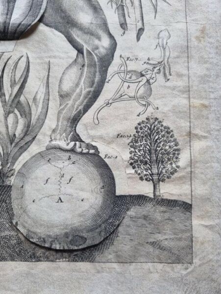
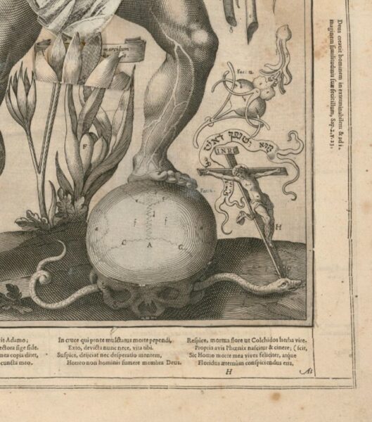
An example of the illustrated details of the original work (left) compared to our later edition (right)
Perhaps having this book in the collection suggests that someone in the Vane family held an interest in anatomy. The wear and tear seen on some of the flaps indicates the book has been well used throughout the years.

